Victor X.D. Yang
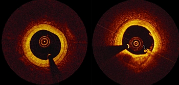
The current standard of care for the assessment of carotid plaques has been the use of ultrasonography, MR imaging, and CT to identify key features of atherosclerosis. Fundamental markers such as intimal morphology, presence of macrophages, and lipid content in the plaque are attributed to the classification of the lesion, as well as assessment of stroke risk. A challenge for the aforementioned imaging modalities is that some of these characteristics are often below their resolution limit. It has been shown that optical coherence tomography (OCT) has the capability of resolving the important markers in the coronary vessels. The application of this optical imaging modality has not fully matured for imaging of carotid arteries.
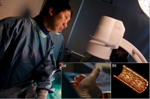
As a result, we are exploring the various potential techniques and applications in preclinical animal carotid models, including nonocclusive saline-based flushing for optimal OCT imaging of vessel wall,1 flow velocity,2 and stent visualization.3 Such techniques reduce contrast load and radiation exposure to patients because optical imaging, as opposed to x-ray based methods, penetrates through saline and the vessel wall to

