Warning: Declaration of My_Walker::start_el(&$output, $item, $depth, $args) should be compatible with Walker_Nav_Menu::start_el(&$output, $data_object, $depth = 0, $args = NULL, $current_object_id = 0) in /home2/ajnrblog/public_html/ajnrdigest/wp-content/themes/ajnr/functions.php on line 258
Classification of High-Grade Glioma into Tumor and Nontumor Components Using Support Vector Machine - AJNR News Digest
March-April 2018
ADULT BRAIN
Figure 2 from Blumenthal

Classification of High-Grade Glioma into Tumor and Nontumor Components Using Support Vector Machine

Dafna Ben Bashat

Moran Artzi

Deborah T. Blumenthal

MRI is the method of choice for the assessment of patients with brain tumors; however, standard MR imaging leaves certain areas indeterminate regarding the diagnosis and evaluation of tumor response. With the rapid introduction of new therapies, specifically for high-grade gliomas, new radiologic patterns have emerged, which challenge radiologic interpretation based on conventional methods alone. Concomitantly, the development of advanced MR methods that enable multiparametric characterization of the tissue further challenge radiologic reading, making it almost impossible to assess by the eye alone. Computer-aided diagnostic tools can enhance the diagnostic capabilities of physicians, reduce the time required for accurate diagnosis, and improve diagnostic and therapy response assessment.

For more than a decade, our group, which includes neuroradiologists, neuro-oncologists, and MR and computer science researchers, has focused on developing CAD tools, utilizing machine-learning algorithms based on advanced and conventional MR methods to improve diagnosis and prognosis and to enable the early prediction of therapy response assessment of patients with brain tumors.

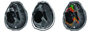
In this study, we attempted to deal with some inherent issues relating to pseudoprogression and pseudoresponse in treatment response assessment of patients with high-grade gliomas by automatically segmenting enhancing and nonenhancing regions into tumor and nontumor tissues.

The current standard for radiologic assessment in patients with high-grade gliomas relies on the Response Assessment in Neuro-Oncology (RANO) criteria, which expand upon the earlier MacDonald criteria, to incorporate the nonenhancing components of tumors, as these components may indicate infiltrative or diffuse tumor growth.

Our proposed method, referred to as segmented RANO (sRANO), provides a logical next step in the evolution of MR clinical imaging to better assess tumor growth and therapy response. Both enhancing and nonenhancing lesion areas may include tumor and nontumor components. The proposed sRANO classifies the lesion areas and defines each component separately. In 16% of our patients followed longitudinally, we were able to identify tumor progression several months in advance of RANO criteria. We envision sRANO to be incorporated into standard MRI assessment for routine follow-up of brain tumors for the prediction of early progression and the response to treatment.

We are currently working on expanding our data base and developing an automated CAD tool to be integrated as a decision-making tool for the clinical assessment and follow-up of patients with high-grade gliomas. We are also investigating the applicability of automatic differentiation between primary brain tumors and metastasis and automatic classification of brain metastases based on their origins. In addition, we are incorporating a radiomics approach, which has gained importance in cancer research in recent years. This approach has been shown to improve prediction over standard radiologic assessments, and was suggested to have an important application in the field of personalized medicine.

Our aim as a group is to develop tools to assist the radiologist and treating clinician, improve diagnosis and follow-up, and enable the accurate and early prediction of therapy response.

Read this article at AJNR.org …