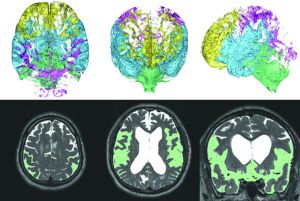
The key feature for the diagnosis of idiopathic NPH is compression of the convexity of the brain, which is concurrent with decreased subarachnoid spaces at the convexity due to z-axial expansion of bilateral ventricles and the Sylvian fissure (ie, disproportionately enlarged subarachnoid space hydrocephalus).
From now on, a lot of radiologists, neurologists, and neurosurgeons may notice how many patients have not been correctly diagnosed with idiopathic NPH.
I was invited to several meetings, including the LANCET Neurology Conference in 2016, the World Congress of Neuroradiology, and XXI Symposium Neuroradiologicum in 2018. Additionally, idiopathic NPH was featured in the Japanese medical journal Clinical Imaging in November 2017. The number of doctors who understand the clinical and radiologic characteristics of the 3 NPH categories (ie, idiopathic NPH, secondary NPH, and adult-onset congenital NPH) is increasing.
Now, I observe not only the 3D distribution of CSF, but also the 3D movement of CSF. Namely, 4D flow is my current topic. I would like to publish our work in the near future.
Every autumn at the annual meeting of the International Society for Hydrocephalus and Cerebrospinal Fluid Disorders, many cutting-edge presentations and exciting discussions take place. We were able to attract the attention of more than 200 experts during the last annual meeting in Kobe, Japan. We are looking forward to this year's Tenth Annual Meeting, which is scheduled to be held October 19–22 in Bologna, Italy.
Read this article at AJNR.org …

