Pierre Lehmann
The importance of diffusion-weighted MR imaging in the detection of recurrent cholesteatoma was already known and quite well documented at the time of this study.
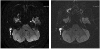
At the beginning, our 3T MRI was mostly used to explore brain. 3T allowed lowering section thickness without signal deterioration or consuming additional time. PROPELLER was used mainly to reduce movement artifacts. We noted fewer artifacts around the temporal bone with DWI sequences. Thanks to the PROPELLER technique, 3T DWI presents better contrast and reduction of artifacts. That was the beginning of our study.
For recurrent cholesteatoma, PROPELLER DWI allows an easier interpretation in two main ways: first, diffusion imaging presents a better contrast than T1-weighted imaging and the cholesteatoma hypersignal is easier to detect; second, PROPELLER DWI reduces artifacts. On a 3T MR imaging unit, artifacts are present much more often than on a 1.5T imaging unit.
