Warning: Declaration of My_Walker::start_el(&$output, $item, $depth, $args) should be compatible with Walker_Nav_Menu::start_el(&$output, $data_object, $depth = 0, $args = NULL, $current_object_id = 0) in /home2/ajnrblog/public_html/ajnrdigest/wp-content/themes/ajnr/functions.php on line 258
Advanced Diffusion Imaging - AJNR News Digest
May 2014
Introduction

Advanced Diffusion Imaging

Jason Allen

Jason Allen

It has been apparent since the time of the early neuroanatomists that the white matter of the CNS was organized in predictable bundles. With the advent of neuroimaging, in particular, MRI, a number of these white matter tracts could be identified. However, the complexity of several of these tracts, particularly their branching and intersecting patterns, limits the utility of conventional MRI techniques. It has also been recognized that the diffusion of water molecules may be restricted by these tightly packed white matter tracts, which led to the development of diffusion tensor imaging (DTI)1 and markedly improved the imaging of the course and integrity of the major white matter tracts in both normal and disease states. With the growing adoption of DTI techniques, two important impediments have been encountered: the problem of multiple intersecting white matter tracts within a single voxel, which limits the ability to trace particular pathways in their entirety, and, on a molecular level, whether current DTI methodology accurately predicts water diffusion.

Conceptually it is easy to understand the difficulty in tracing white matter tracts when there is intravoxel fiber crossing between two or more different bundles. Multiple data-acquisition and computational models have been developed to address this problem. High angular resolution diffusion imaging (HARDI) involves the acquisition of a large number of gradient directions and, in a simplified form, all possible fiber directions are considered together during analysis with the path with the largest consistency in neighboring voxel direction identified as the most probable connection and, hence, the most likely direction for the fiber tract under consideration.2 Traditional DTI methodology tends to provide erroneous tract termination or directions when encountering complex intravoxel fiber crossing, whereas HARDI provides a more accurate representation of the underlying white matter architecture.

The development of diffusion kurtosis imaging (DKI) has provided a potentially more fundamental change in the approach to water diffusion imaging. Molecular diffusion is a random process and may be depicted by a probability distribution. If the substrate through which a water molecule is traversing is homogeneous, its distribution will have a Gaussian form, with the width of the distribution proportional to the apparent diffusion coefficient as is assumed in DTI methodology. However, water diffusion within complex biological substrates, which typically contain multiple tissue types and compartments, does not adhere to a Gaussian distribution. Kurtosis is a dimensionless metric that describes the deviation of a probability distribution from a Gaussian form and methods have been developed to estimate the kurtosis of water diffusion within the CNS.3 DKI is an extension of DTI and is acquired similarly, with the exception of the addition of higher b-values, and the same metrics of fractional anisotropy (FA) and mean diffusivity (MD) may be computed. Mean kurtosis (MK) is an additional value that may be calculated using DKI, which, again, is the measure of the degree to which water diffusion departs from a Gaussian distribution in a particular region. In the most straightforward analysis, DKI may more accurately characterize white matter tracts than DTI, possibly allowing better tracking of intersecting pathways. As MK may be conceptualized as a measure of diffusional heterogeneity of a particular region, a possible surrogate marker for the inherent complexity of a region, DKI may also provide better characterization of both gray and white matter.

The current issue of the AJNR News Digest highlights advanced MRI diffusion imaging methods, including the application of HARDI in the mapping of the complex auditory radiation pathway and the use of DKI in the investigation of diverse disease states such as Alzheimer disease (AD), multiple sclerosis, and squamous cell carcinoma as well as in the characterization of the developing brain.

In the first article, Berman et al demonstrate the superiority of HARDI to traditional DTI in the delineation of the auditory radiation, which has been previously difficult to track with imaging.4 As discussed above, traditional DTI has limited utility in situations of significant intravoxel fiber crossing or complexity. The robust anteroposteriorly aligned inferior longitudinal fasciculus (ILF) traverses the auditory radiation and DTI fiber tracking is
generally overwhelmed by the dominant ILF and only depicts that tract; however, as demonstrated by Berman et al, HARDI is capable of depicting the auditory radiation despite this crossing fiber tract. This may allow research into the disruption of the auditory radiation in various conditions and also provides further evidence of the superiority of the HARDI technique.

The next article utilizes DKI in the investigation of cognitive dysfunction due to AD. Fieremans et al apply DKI methodology in the assessment of AD-related white matter microstructural changes.5 Their paper demonstrates several DKI-derived white matter metrics, in particular extra-axonal space diffusivity and axonal water fraction, which distinguish between healthy controls, patients with amnestic mild cognitive impairment, and patients with AD.

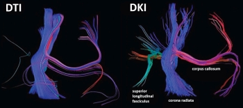
In the article by Paydar et al, DKI is compared with DTI in measuring age-related white and gray matter microstructural changes in the developing human brain.6 This study is particularly interesting in that DKI metrics, in particular, MK, continue to increase past the plateau for DTI values in both white and gray matter. For example, 90% of maximal FA was attained by 5 months of age within the external capsule, whereas this level was not reached until 18 months of age for MK. Their data provide further support for the theory that MK may reflect underlying tissue heterogeneity, which increases during development, particularly in gray matter. Finally, Paydar et al demonstrate that, similar to HARDI, DKI tractography may be better able to accurately depict white matter architecture in areas of complex intravoxel fiber crossing.

Raz et al apply DKI in conjunction with traditional DTI methods to characterize both white and gray matter spinal cord pathology in patients with multiple sclerosis (MS).7 This paper confirms previously demonstrated white matter changes such as reduced FA and increased MD and also demonstrates decreased MK in gray matter within the spinal cord. Raz et al suggest that the decreased gray matter MK, which reflects reduced diffusional heterogeneity, may be associated with irreversible degenerative changes and disability in MS.

In the final article, Jansen et al utilize DKI to investigate patients with head and neck squamous cell carcinomas.8 This feasibility study demonstrates the possible utility of DKI in regions outside of the CNS and also raises the potential utility of DKI-derived metrics, including the apparent kurtosis coefficient, in the further characterization of head and neck cancers.

As more investigators adopt advanced MRI techniques beyond traditional DTI methodology, it is likely that the popularity of both HARDI and DKI will continue to increase. Both of these methods address a limitation of conventional MRI techniques, intravoxel heterogeneity/complexity, which is a fundamental property of the CNS that may also be a marker of dysfunction when disrupted.

References

  1. Basser PJ, Jones DK. Diffusion-tensor MRI: theory, experimental design and data analysis—a technical review. NMR Biomed 2002;15:456–67, 10.1002/nbm.783
  2. Chung HW, Chou MC, Chen CY. Principles and limitations of computational algorithms in clinical diffusion tensor MR tractography. AJNR Am J Neuroradiol 2011;32:3–13, 10.3174/ajnr.A2041
  3. Jensen JH, Helpern JA, Ramani A, et al. Diffusional kurtosis imaging: the quantification of non-Gaussian water diffusion by means of magnetic resonance imaging. Magn Res Med 2005;53:1432–40, 10.1002/mrm.20508
  4. Berman JI, Lanza MR, Blaskey L, et al. High angular resolution diffusion imaging probabilistic tractography of the auditory radiation. AJNR Am J Neuroradiol 2013;34:1573–78, 10.3174/ajnr.A3471
  5. Fieremans E, Benitez A, Jensen JH, et al. Novel white matter tract integrity metrics sensitive to Alzheimer disease progression. AJNR Am J Neuroradiol 2013;34:2105–12, 10.3174/ajnr.A3471
  6. Paydar A, Fieremans E, Nwankwo JI, et al. Diffusional kurtosis imaging of the developing brain. AJNR Am J Neuroradiol 2013 [Epub ahead of print], 10.3174/ajnr.A3764
  7. Raz E, Bester M, Sigmund EE, et al. A better characterization of spinal cord damage in multiple sclerosis: a diffusional kurtosis imaging study. AJNR Am J Neuroradiol 2013;34:1846–52, 10.3174/ajnr.A3512
  8. Jansen JFA, Stambuk HE, Koutcher JA, et al. Non-Gaussian analysis of diffusion-weighted MR imaging in head and neck squamous cell carcinoma: a feasibility study. AJNR Am J Neuroradiol 2010;31:741–48, 10.3174/ajnr.A1919


Image modified from: Paydar A, Fieremans E, Nwankwo JI, et al. Diffusional Kurtosis Imaging of the Developing Brain.