Orit Glenn
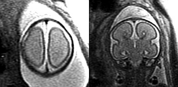
MRI is increasingly used to evaluate the fetal brain in cases where an abnormality is detected by routine prenatal sonography and in cases where the fetus is at increased risk for abnormalities in brain development. As such, it allows us to diagnose brain abnormalities in utero that are often missed by prenatal ultrasound.
I chose to study agenesis of the corpus callosum in fetuses using MRI because of my interest in the developing fetal brain and because of the challenges in prenatal counseling of fetal callosal agenesis. I also chose to study this topic because more radiologists are performing fetal MRI, and it is important for us to be familiar with the types of MRI findings that can be seen in fetuses with agenesis of the corpus callosum.
The results of this study have influenced my clinical practice in that I always carefully evaluate the fetal brain for additional abnormalities when callosal agenesis is identified on fetal MRI. In particular, I look carefully for abnormalities of sulcation, brain stem, and cerebellum on fetal MRI. Moreover, this study demonstrates that most of the additional abnormalities detected by fetal MRI in fetuses with callosal agenesis are missed on prenatal ultrasound. The results of this study have also influenced how our
