
Sato Eida
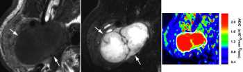
Preoperative prediction of tumor malignancy is clinically very important, because this information strongly influences the surgical plan. Diffusion-weighted imaging is very sensitive to biophysical abnormalities associated with pathologic changes; in this regard, apparent diffusion coefficient measurement has been used to diagnose benign and malignant salivary gland tumors. However, salivary gland tumors are composed of distinctive tissues, including proliferating tumor cells, myxomatous tissues, lymphoid tissues, necrosis, and cysts. Analyzing a large region of interest in a histologically heterogeneous tumor may therefore result in spurious results with regard to the tumor histology. To avoid this potential error, tissue characterization using high-resolution MRI techniques is mandatory. In this study, we reasoned that the ADC mapping technique might enable effective characterization of the histologic features of salivary gland tumors. The ADC maps showed that a greater number of areas with high ADCs (≧1.8 × 10⁻³mm2/s) were significantly greater in benign tumors than in malignant tumors. The sensitivity and specificity for high ADC occupying fewer than 5% of the tumor area was 89% and 100%, respectively, resulting in 97% accuracy, 100% positive predictive value, and 96% negative predictive value. The results of our study suggest the feasibility of an ADC map in differentiating between benign and malignant diseases in the head and neck region.