Warning: Declaration of My_Walker::start_el(&$output, $item, $depth, $args) should be compatible with Walker_Nav_Menu::start_el(&$output, $data_object, $depth = 0, $args = NULL, $current_object_id = 0) in /home2/ajnrblog/public_html/ajnrdigest/wp-content/themes/ajnr/functions.php on line 258
Development of High Signal Intensity within the Globus Pallidus and Dentate Nucleus following Multiple Administrations of Gadobenate Dimeglumine - AJNR News Digest
November-December 2019
ADULT BRAIN
Figure 2 from Bolles

Development of High Signal Intensity within the Globus Pallidus and Dentate Nucleus following Multiple Administrations of Gadobenate Dimeglumine

Roberts picture

Donna R. Roberts

Bolles picture

Genevieve M. Bolles

Gadolinium-based contrast agents (GBCAs) have been used routinely in neuroradiology since their introduction in the late 1980s and provide invaluable clinical information for the diagnosis and management of patients with neurologic diseases. These agents have been generally considered safe for patients with normal renal function, with over 450 million doses administered worldwide.

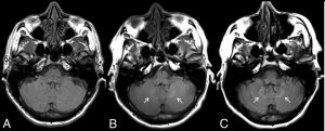
In 2014, Kanda et al1 reported an association between the number of prior GBCA administrations and the development of hyperintense signal in the globus pallidus and dentate nucleus on unenhanced T1-weighted images. Shortly afterwards, McDonald et al2 confirmed the presence of gadolinium within brain tissue in adult decedents who had undergone GBCA-enhanced MRI. We have shown on MRI and at autopsy that gadolinium is also retained within the pediatric brain.3–5

GBCAs can be classified as either macrocyclic or linear based on the structure of the gadolinium chelate. Several studies, in both adult and pediatric populations, have evaluated various GBCAs with the general consensus being that linear, but not macrocyclic, GBCAs are associated with long-term gadolinium retention in the brain. In our study, we found the development of high signal intensity within the globus pallidus and dentate nucleus following multiple administrations of the linear agent gadobenate dimeglumine.6

Currently, there is little scientific evidence linking gadolinium deposition in the brain with specific clinical symptoms, though this is an area of active research. Our group in particular is studying gadolinium deposition in pediatric patients given the unique vulnerabilities of children, including ongoing central nervous system development, immature renal function, and active bone formation. In adults, gadolinium is known to accumulate in bone tissue at levels approximately 23 times higher than those in the brain.7 It is unknown whether rapid bone turnover due to growth and development enhances gadolinium accumulation in children. To investigate this, we are currently measuring gadolinium levels in sternal bone in children who have undergone cardiac surgery. Results from this study will be important in determining whether bone represents a long-term storage site for gadolinium in children. If so, patients receiving large cumulative GBCA doses during childhood may be at risk for secondary endogenous gadolinium exposure much later in life (eg, during periods of bone loss).

Given the current uncertainties concerning the clinical significance of gadolinium deposition within the body, many practices have chosen to take a cautious stance and have switched to the exclusive use of higher-stability, macrocyclic agents.8 In 2016, the American College of Radiology and the American Society of Neuroradiology issued a joint position statement recognizing the propensity of GBCAs to deposit in sensitive tissues such as the brain.9 In July 2017, the Pharmacovigilance Risk Assessment Committee of the European Medicines Agency recommended suspension of the marketing authorizations for 4 linear GBCAs.10 The U.S. Food and Drug

Administration (FDA) required GBCA manufacturers to add a warning for gadolinium retention to their product labeling in 2017.11 The FDA also requested manufacturers to conduct additional studies to assess the safety of GBCAs.11

As we await the results of long-term research studies to address any potential clinical significance of gadolinium deposition in the body, we should continue to carefully select patients to undergo GBCA-enhanced MRI. The retention characteristics of agents should be considered when choosing a GBCA, and consideration should be given to the use of higher-stability agents, particularly for patients requiring multiple lifetime GBCA doses. Importantly, for appropriate clinical indications, the benefit of GBCA administration continues to outweigh any as-yet-unknown potential risks; therefore, patients should not be deprived of a well-indicated contrasted MR imaging exam.

References

  1. Kanda T, Ishii K, Kawaguchi H, et al. High signal intensity in the dentate nucleus and globus pallidus on unenhanced T1-weighted MR images: relationship with increasing cumulative dose of a gadolinium-based contrast material. Radiology 2014;270:834–41, 10.1148/radiol.13131669
  2. McDonald RJ, McDonald JS, Kallmes DF, et al. Intracranial gadolinium deposition after contrast-enhanced MR imaging. Radiology 2015;275:772–82, 10.1148/radiol.15150025.
  3. Roberts DR, Chatterjee AR, Yazdani M, et al. Pediatric patients demonstrate progressive T1-weighted hyperintensity in the dentate nucleus following multiple doses of gadolinium-based contrast agent. AJNR Am J Neuroradiol 2016;37:2340–47, 10.3174/ajnr.A4891.
  4. Roberts DR, Holden KR. Progressive increase of T1 signal intensity in the dentate nucleus and globus pallidus on unenhanced T1-weighted MR images in the pediatric brain exposed to multiple doses of gadolinium contrast. Brain Dev 2016;38:331–36, 10.1016/j.braindev.2015.08.009.
  5. Roberts DR, Welsh CA, LeBel DP II, et al. Distribution map of gadolinium deposition within the cerebellum following GBCA administration. Neurology 2017;88:1206–08, 10.1212/WNL.0000000000003735.
  6. Bolles GM, Yazdani M, Stalcup ST, et al. Development of high signal intensity within the globus pallidus and dentate nucleus following multiple administrations of gadobenate dimeglumine. AJNR Am J Neuroradiol 2018;39:415–20, 10.3174/ajnr.A5510.
  7. Murata N, Gonzalez-Cuyar LF, Murata K, et al. Macrocyclic and other non-group 1 gadolinium contrast agents deposit low levels of gadolinium in brain and bone tissue: preliminary results from 9 patients with normal renal function. Invest Radiol 2016;51:447–53, 10.1097/RLI.0000000000000252.
  8. Mithal LB, Patel PS, Mithal D, et al. Use of gadolinium-based magnetic resonance imaging contrast agents and awareness of brain gadolinium deposition among pediatric providers in North America. Pediatr Radiol 2017;47:657–64, 10.1007/s00247-017-3810-4.
  9. ACR–ASNR position statement on the use of gadolinium contrast agents [press release]. May 2016. Available at: https://www.asnr.org/wp-content/uploads/2017/03/ACR_ASNR_Position_Statement_on_the_Use_of_Gadolinium_Contrast_Agents.pdf. Accessed September 24, 2019.
  10. PRAC concludes assessment of gadolinium agents used in body scans and recommends regulatory actions, including suspension for some marketing authorisations [press release]. October 3, 2017. Available at: http://www.ema.europa.eu/ema/index.jsp?curl=pages/news_and_events/news/2017/03/news_detail_002708.jsp&mid=WC0b01ac058004d5c1. Accessed September 24, 2019.
  11. FDA Drug Safety Communication: FDA warns that gadolinium-based contrast agents (GBCAs) are retained in the body; requires new class warnings [press release]. December 19, 2017. Available at: https://www.fda.gov/Drugs/DrugSafety/ucm589213.htm Accessed September 24, 2019.

Read this article at AJNR.org …