Warning: Declaration of My_Walker::start_el(&$output, $item, $depth, $args) should be compatible with Walker_Nav_Menu::start_el(&$output, $data_object, $depth = 0, $args = NULL, $current_object_id = 0) in /home2/ajnrblog/public_html/ajnrdigest/wp-content/themes/ajnr/functions.php on line 258
Diffusional Kurtosis Imaging of the Developing Brain - AJNR News Digest
May 2014
Pediatrics

Diffusional Kurtosis Imaging of the Developing Brain

Amir Paydar

Amir Paydar

Human embryology and development is indeed one of the most (if not the most) fascinating subjects in the field of biological sciences. Nevertheless, our scientific community still faces challenges in trying to understand the concepts that define the underlying mechanisms of embryology and tissue development. After all, it is a very complex subject to grasp and many of the processes that take place from zygote to adulthood are yet to be ascertained. Despite this challenge, we have come to recognize that understanding the natural course of normal tissue development on both microscopic and macroscopic scales is the key to deciphering the mechanisms that take place during tissue injury and degeneration as well as healing and scarring.

Realizing this concept, my good friends and colleagues at New York University (NYU) Medical Center decided to take on an ambitious human study to investigate brain maturation using noninvasive imaging techniques in the pediatric population. Our research subjects included 59 healthy infants with an age spectrum ranging from birth to approximately 5 years of age, when the postnatal brain is in its most active stage of development. We implemented an innovative MRI diffusion technique called diffusional kurtosis imaging (DKI) to study the microstructural changes that occur in both the WM and GM in the developing brain.

Macrostructural changes that take place within the brain during the course of maturation have been well documented by conventional MRI techniques in both normal and pathologic states. However, these conventional techniques are limited in their ability to quantify developmental changes that occur at the microstructural level. Diffusion imaging, particularly the widely used diffusion tensor imaging (DTI), has been shown to be sensitive to age-related microstructural changes that occur within the network of WM tracks as a result of myelination. However, DTI is based on
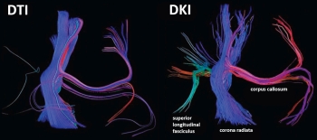
a Gaussian approximation of water diffusion, which limits its sensitivity to diffusional and microstructural properties of biological tissues. On the other hand, the recently developed DKI technique, which is a clinically feasible extension of the traditional DTI model, takes into account the non-Gaussian diffusional properties of water motion in complex media and is therefore more comprehensive in evaluating brain tissue microstructural complexity.

In our study, we hypothesized that, owing to its potentially higher sensitivity for detection of age-related microstructural changes, DKI may provide additional information about brain maturation when compared with that obtainable with conventional DTI in both WM and GM. Our results were quite conclusive and we received tremendous positive feedback after presenting and publishing our findings.

Our data revealed that both DTI and DKI can reflect the age-related increase in diffusional anisotropy in WM tracts, predominantly as a result of myelination and axonal packing, during the first 2 years of life. However, compared to DTI, DKI offers additional characterization of the isotropic diffusion barriers that continue to develop in the WM beyond the first 2 years (ie, after myelination has already peaked). DKI can also better resolve the progression of GM organization with respect to age by accounting for other isotropic microstructural barriers that form at the cellular level in the GM. Accordingly, DKI is an innovative non-Gaussian diffusion MRI technique that offers more sensitive and comprehensive measures for the quantitative evaluation of age-related changes in the microstructural complexity of both WM and GM.

In summary, DKI is an excellent noninvasive technique for quantifying microstructural changes in the brain. But what is the potential clinical utility of this discovery? We know that the diffusion barriers that form due to the progressive increase in macromolecular reorganization during brain maturation can be altered in the settings of tissue injury, degeneration, and healing. Therefore, DKI may potentially serve as a valuable measuring tool for detection of pathologic cellular processes that affect microstructural changes in the developing WM and GM. Moreover, DKI may allow quantitative evaluation of pathologies that alter microstructural complexity in the adult brain as well. We are optimistic about this great prospect and hope that the neuroscience/neuroradiology community will join our research team at NYU in effectively using this innovative MRI diffusion technique for both research and clinical purposes in the near future.


Read this article at AJNR.org . . .