Tobias Struffert
Endovascular treatment of ischemic stroke has developed rapidly in recent years. I performed my first mechanical stroke treatment with a dedicated stroke device in a patient included in the penumbra trial that was published in AJNR in 2008. This patient showed a dramatic improvement of his clinical condition right after recanalization of the left MCA. Of course, improvement of these techniques was a subject of our further efforts.
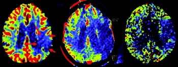
It is obvious and shown by many studies that we can recanalize occluded intracranial vessels within a short time. On the other hand, we have to admit that in a high percentage of cases the patients’ clinical condition may not improve despite recanalization. So why should we try to perform stroke imaging within the angio suite? Because we can save time and select patients. Outcomes are highly correlated with time from stroke onset to reperfusion. Perfusion imaging may allow us to distinguish those patients most likely to benefit from reperfusion from those where mechanical thrombectomy may not result in a good outcome, and may possibly add risk due to reperfusion trauma. Enabling diagnosis, selection, and treatment within an identical environment will improve and accelerate workflow. Additionally, monitoring during treatment becomes feasible.
This new method is challenging. The value of perfusion imaging is not yet fully understood and still under review. We now have this tool available at a point in time and location in which it has never been available before. This will require further analysis to understand the predictive value of perfusion measurements.
