Warning: Declaration of My_Walker::start_el(&$output, $item, $depth, $args) should be compatible with Walker_Nav_Menu::start_el(&$output, $data_object, $depth = 0, $args = NULL, $current_object_id = 0) in /home2/ajnrblog/public_html/ajnrdigest/wp-content/themes/ajnr/functions.php on line 258
Human Papillomavirus–Associated Head & Neck Cancer - AJNR News Digest
June 2015
Introduction

Human Papillomavirus–Associated Head & Neck Cancer

Guest Editor
Ryan T. Fitzgerald

Human papillomavirus (HPV) is now recognized as the primary causative factor in a subgroup of epidemiologically and clinically distinct cancers of the head and neck, oropharyngeal squamous cell carcinoma (OPSCC).1,2 The rising incidence of OPSCC relative to squamous cell cancers elsewhere within the mucosal spaces of the head and neck is thought to be directly related to the predilection of the HPV-associated disease for the oropharynx and increasing proportion of head and neck cancers that are HPV-positive. The prevalence of HPV positivity among oropharyngeal tumors has increased from under 17% during the 1980s to almost 72% during the 2000s.1 Compared with non-HPV OPSCC, HPV-associated OPSCC is more commonly diagnosed at a younger age. Over time, the age at which HPV-associated OPSCC is diagnosed is decreasing on the order of 0.5 years per decade from 1973 through 2004.3 Human papillomavirus subtype 16 (HPV-16) is by far the most common virus detected in OPSCC specimens, accounting for approximately 95% of the disease burden from among 37 viral subtypes.4 Both of the FDA approved HPV vaccines, Cervarix (Glaxo Smith Kline, Brentford, England) and Gardasil (Merck & Co., Kenilworth, NJ), provide coverage for HPV-16.

Risk factor profiles for HPV and non-HPV OPSCC differ considerably. Associations with HPV-16-positive head and neck squamous cell carcinoma (HNSCC) include the number of oral sex partners and marijuana use (including intensity, duration, and cumulative joint-years). In contrast, HPV-16-negative HNSCC is strongly associated with tobacco smoking, alcohol use, and poor oral hygiene.4 HPV-16-negative HNSCC has not been shown to correlate with prior sexual behavior or marijuana use.4 Such divergent risk profiles support viewing HPV-positive and -negative cancers as distinct clinicopathologic entities. Interestingly, cannabinoids are known to possess immunosuppressant effects that may potentially contribute to the role of marijuana in the development of HPV-induced HNSCC through increased risk of infection upon exposure, promoting persistence of HPV infection long-term, and decreased antineoplastic immune surveillance.4

Tumor HPV status is recognized as a robust and independent prognostic indicator for improved survival among patients with OPSCC. In a 2010 study, 3-year survival of patients with OPSCC whose tumors were HPV-positive, after adjustment for age, race, tumor/nodal stage, tobacco exposure, and treatment assignment, was 82% vs 57% for patients with HPV-negative tumors.5 Such a survival benefit is thought to be related at least in part to higher intrinsic sensitivity of HPV-positive tumors to radiation and/or to increased radiosensitization with the use of combined radiation and cisplatin. Five-year survival among patients with HPV-positive tumors approaches 80% versus 45–50% among those with HPV-negative tumors. Further, the HPV-associated survival benefit has been shown to persist beyond 15 years after initial diagnosis, as demonstrated in a study of 271 patients with OPSCC from 1984–2004 that showed median survival of 131 months in HPV-positive cancer compared with 20 months for the HPV-negative cohort.3
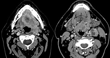
This month’s AJNR Digest highlights 5 recent articles from the journal that examine various facets of the imaging approach for diagnosis and surveillance of HPV-related OPSCC. Cantrell et al describe differences in the CT imaging phenotype between HPV-positive and HPV-negative disease, with HPV-positive lesions more commonly displaying well-defined borders and cystic nodal metastases.6 Buch et al report on a CT-based texture analysis method that showed statistically significant differences between HPV-positive and -negative lesions and thus represents an image-based tool that shows promise for the prediction of HPV status of oropharyngeal squamous cell carcinomas in vivo.7 Matoba et al provide a report on the use of diffusion-weighted imaging as a biomarker for predicting treatment response in HNSCC treated with chemoradiotherapy.8 Pickering et al further explore the link between imaging features and tumor genomics of cancers occurring in the oral cavity.9 And lastly, Hamilton et al introduce a composite scoring system for persistent nodal metastasis in patients with OPSCC that yielded improved specificity relative to established criteria while preserving sensitivity.10

References

  1. Chaturvedi AK, Engels EA, Pfeiffer RM, et al. Human papillomavirus and rising oropharyngeal cancer incidence in the United States. J Clin Oncol 2011;29:4294–4301, 10.1200/JCO.2011.36.4596
  2. Vainshtein J, McHugh JB, Spector ME, et al. Human papillomavirus-related oropharyngeal cancer: HPV and p16 status in the recurrent versus parent tumor. Head Neck 2014;37:8–11, 10.1002/hed.23548
  3. Chaturvedi AK, Engels EA, Anderson WF, et al. Incidence trends for human papillomavirus-related and -unrelated oral squamous cell carcinomas in the United States. Journal of Clinical Oncology 2008;26:612–19, 10.1200/JCO.2007.14.1713
  4. Gillison ML, D'Souza G, Westra W, et al. Distinct risk factor profiles for human papillomavirus type 16-positive and human papillomavirus type 16-negative head and neck cancers. J Natl Cancer Inst 2008;100:407–20, 10.1093/jnci/djn025
  5. Ang KK, Harris J, Wheeler R, et al. Human papillomavirus and survival of patients with oropharyngeal cancer. N Engl J Med 2010;363:24–35, 10.1056/NEJMoa0912217
  6. Cantrell SC, Peck BW, Li G, et al. Differences in imaging characteristics of HPV-positive and HPV-negative oropharyngeal cancers: a blinded matched-pair analysisAJNR Am J Neuroradiol 2013;34:2005–09, 10.3174/ajnr.A3524
  7. Buch K, Fujita A, Li B, et al. Using texture analysis to determine human papillomavirus status of oropharyngeal squamous cell carcinomas on CT. AJNR Am J Neuroradiol published ahead of print April 2, 2015, 10.3174/ajnr.A4285
  8. Matoba M, Tuji H, Shimode Y, et al. Fractional change in apparent diffusion coefficient as an imaging biomarker for predicting treatment response in head and neck cancer with chemoradiotherapy. AJNR Am J Neuroradiol 2014;35:379–85, 10.3174/ajnr.A3706
  9. Pickering CR, Shah K, Ahmed S, et al. CT imaging correlates of genomic expression for oral cavity squamous cell carcinoma. AJNR Am J Neuroradiol 2013;34:1818–22, 10.3174/ajnr.A3635
  10. Hamilton JD, Ahmed S, Sandulache VC, et al. Improving imaging diagnosis of persistent nodal metastases after definitive therapy for oropharyngeal carcinoma: specific signs for CT and best performance of combined criteriaAJNR Am J Neuroradiol 2013;34:1637–42, 10.3174/ajnr.A3461

Image modified from: Cantrell SC, Peck BW, Li G, et al. Differences in imaging characteristics of HPV-positive and HPV-negative oropharyngeal cancers: a blinded matched-pair analysis.