Warning: Declaration of My_Walker::start_el(&$output, $item, $depth, $args) should be compatible with Walker_Nav_Menu::start_el(&$output, $data_object, $depth = 0, $args = NULL, $current_object_id = 0) in /home2/ajnrblog/public_html/ajnrdigest/wp-content/themes/ajnr/functions.php on line 258
May-June 2019 Edition of AJNR's News Digest: Inner Ear Malformations
May-June 2019
Introduction

Inner Ear Malformations: More to Come?

Suresh Mukherji

Inner ear malformations are one of the most challenging and confusing topics in all of neuroradiology (and radiology for that matter). The topic has “haunted” radiology residents for years, and I remember being a petrified fourth-year resident in the spring of 1992 (last century!), preparing for the oral boards and being completely flummoxed by the temporal bone. I remember asking one of my neuroradiology faculty about how to approach the temporal bone for the oral boards. His answer was, “If they show you a weird temporal bone CT, just say Mondini malformation and go to the next case!” Based on my conversations with many colleagues since that time, I believe many adopted this strategy!

The most widely accepted approach to categorizing inner ear malformations for many years was published by Jackler et al1 in 1987. It was a very practical approach that was based on the embryonic development of the inner structures. The inner malformations were categorized according to when the arrest in fetal development occurred. The most “benign” of these malformations is the abnormality reported by Mondini in his seminal 1791 article, which occurs at a late stage in inner ear development.2–4

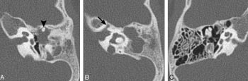
Technical improvements in CT and MRI have resulted in our ability to visualize detailed structures of the inner ear that previously could only be identified in histology textbooks. Lemmerling et al5 and Davidson et al6 published 2 very important papers in the late 1990s that identified subtle malformations of the modiolus causing subtle contour abnormalities of the cochlea in patients with enlarged vestibular aqueducts. These important works were the first to describe modiolar malformations previously documented by Schuknecht in his classic textbook.7

Sennaroglu and Saatci8 introduced their classification of inner ear malformations in 2002. Their approach was a modification of Jackler’s classification but introduced the term “cystic cochleovestibular malformation” to describe an inner ear malformation that had an incompletely formed cochlea and vestibule. This malformation was also referred to as “incomplete partition type I (IP-I).” The classic Mondini malformation was called “incomplete partition type II (IP-II).”

This edition of the AJNR News Digest contains several important articles that review the current classification of inner ear malformations, identify ways to detect subtle cochlear malformations, highlight potential genetic associations with inner ear anomalies, and highlight the role of 7T MRI in furthering our understanding of the normal anatomy and subtle malformations.

Huang et al9,10 present a state-of-the-art review of sensorineural hearing loss and review the current Sennaroglu classification of inner ear malformations. Booth et al11 and Reinshagen et al12 expand the seminal findings reported by Lemmerling et al and Davidson et al and describe different measurements to detect subtle cochlear IP-II malformations that could otherwise be easily overlooked. The article by Verheij et al13 discusses the anatomic malformations associated with 22q11.2 deletion syndrome.

This important article reminds us that inner ear malformations cannot solely be attributed to an arrest in embryogenesis and that genetic mutations may play an important role in inner ear malformations, especially those malformations that cannot be explained by arrested development. Finally, the article by van der Jagt et al14 provides us an exciting glimpse of the potential future of temporal bone imaging at 7T with the ability to visualize the scala media.

These outstanding articles highlight the continuing advancements in imaging that are improving our understanding of the intricate normal anatomy of the inner ear, which previously could only be identified in histologic specimens. We are now detecting subtle anomalies that were only visible in pathology textbooks. I am quite sure this powerful convergence will eventually result in a new classification of inner ear malformations based on congenital anomalies that are yet to be discovered.

References

  1. Jackler RK, Luxford WM, House WF. Congenital malformations of the inner ear: a classification based on embryogenesisLaryngoscope1987;97(S40):1–14, 10.1002/lary.5540971301.
  2. Mondini C. Anatomia surdi nati sectio: de Bononiensi Scientiarum et Artium Institute atque Academia commentariiBononiae 1791;7:419–28.
  3. Mondini C. Minor works of Carlo Mondini: the anatomic section of a boy born deafAm J Otol1997;18:288–93.
  4. Lo WM. What is a Mondini and what difference does a name make? AJNR Am J Neuroradiol1999;20:1442–44.
  5. Lemmerling MM, Mancuso AA, Antonelli PJ, et al. Normal modiolus: CT appearance in patients with a large vestibular aqueductRadiology1997;204:213–19, 10.1148/radiology.204.1.9205250.
  6. Davidson HC, Harnsberger HR, Lemmerling MM, et al. MR evaluation of vestibular cochlear anomalies associated with endolymphatic duct and sacRadiology1999;20:1435–41.
  7. Schuknecht HF. Pathology of the Ear. Cambridge: Harvard University Press; 1974.
  8. Sennaroglu L, Saatchi I. A new classification for cochleovestibular malformationsLaryngoscope2002;112:213–19, 10.1097/00005537-200212000-00019.
  9. Huang BY, Zdanski C, Castillo M. Pediatric sensorineural hearing loss, part 1: practical aspects for neuroradiologistsAJNR Am J Neuroradiol2012;33:211–17, 10.3174/ajnr.A2498.
  10. Huang BY, Zdanski C, Castillo M. Pediatric sensorineural hearing loss, part 2: syndromic and acquired casesAJNR Am J Neuroradiol 2012;33:399–406, 10.3174/ajnr.A2499.
  11. Booth TN, Wick C, Clarke R, et al. Evaluation of the normal cochlear second interscalar ridge angle and depth on 3D T2-weghted images: a tool for the diagnosis of scala communis and incomplete partition type IIAJNR Am J Neuroradiol2018;39:923–27, 10.3174/ajnr.A5585.
  12. Reinshagen KL, Curtin HD, Quesnel AM, et al. Measurement for detection of incomplete partition type II anomalies on MR imagingAJNR Am J Neuroradiol2017;38:2003–07, 10.3174/ajnr.A5335.
  13. Verheij E, Elden L, Crowley TB, et al. Anatomic malformations of the middle and inner ear in 22q11.2 deletion syndrome: case series and literature reviewAJNR Am J Neuroradiol2018;39:928–34, 10.3174/ajnr.A5588.
  14. van der Jagt MA, Brink WM, Versluis MJ, et al. Visualization of human inner ear anatomy with high resolution MR at 7T: initial clinical assessmentAJNR Am J Neuroradiol2015;36:378–83, 10.3174/ajnr.A4084.