Warning: Declaration of My_Walker::start_el(&$output, $item, $depth, $args) should be compatible with Walker_Nav_Menu::start_el(&$output, $data_object, $depth = 0, $args = NULL, $current_object_id = 0) in /home2/ajnrblog/public_html/ajnrdigest/wp-content/themes/ajnr/functions.php on line 258
Intracranial Hypotension: Diagnosis and Treatment - AJNR News Digest
October 2013
Introduction

Intracranial Hypotension: Diagnosis and Treatment

Jeffrey Ross

Jeffrey Ross

The Agony of Aliquorrhea

Intracranial hypotension can occur in a variety of clinical settings and can be grouped by primary or secondary etiologies. We are generally quite familiar with the secondary type of intracranial hypotension, related to prior cranial or spinal surgery, trauma, or prior lumbar puncture, as a cause of postural headache. The primary form (aka spontaneous intracranial hypotension) has now received sufficient attention in the literature that this diagnosis should not be missed or misinterpreted.1,2 Nevertheless, it can present as a therapeutic and, to a lesser extent, diagnostic dilemma.

The seemingly varied signs present on cranial and spinal imaging can be explained by the Monro-Kellie doctrine, which states that, with an intact skull, the sum of the volume of brain + CSF + blood is constant.3 Any increase in one of these three factors must be accompanied by a decrease in one or both of the other factors. Regarding intracranial imaging, this is manifested by 1) brain sagging, 2) engorgement of venous structures, and 3) extra-axial fluid collections. Brain sagging (slumping) is shown by flattening of the ventral pons against the clivus, inferiorly displaced third ventricle, low-lying cerebellar tonsils, loss of the basal cisterns, decreased mamillopontine distance, and draping of the chiasm over the pituitary. Venous engorgement is demonstrated by prominent, rounded dural sinuses, pachymeningeal thickening and enhancement, and by pituitary enlargement. Extra-axial collections (subdural effusions and hematomas) may occur when these compensatory mechanisms are inadequate.

Spine changes are slightly more protean. No direct correlation related to brain sagging is seen in the spine. Any sagging that occurs in the spine relates to the loss of turgor of the thecal sac, with a correspondingly enlarged epidural space that often contains fluid and outlines the exiting nerves. Engorgement of the venous structures is present, with thickened and enhancing dura and prominence of the epidural plexus (which in the cervical spine can be quite alarming in appearance). Spinal subdural effusions—or, less commonly, hematomas—may also occur.

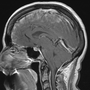
Sagittal T1-weighted postcontrast image shows the typical findings of intracranial hypotension, including low tonsils, flattened ventral pons, decreased mamillopontine distance, plump sella, and dilated intracranial and upper cervical spine veins.

Sagittal T1-weighted postcontrast image shows the typical findings of intracranial hypotension, including low tonsils, flattened ventral pons, decreased mamillopontine distance, plump sella, and dilated intracranial and upper cervical spine veins.

In this issue of the AJNR News Digest, we focus on the work of authors who refined the pathophysiology, imaging patterns, use of advanced imaging techniques, and treatment of intracranial hypotension and CSF leaks.

Concerning the diagnosis of intracranial hypotension, Farb et al4 point us to the utility of the venous distension sign. This method is easily applied, qualitative, not cumbersome in nature, and can be evaluated in all sagittal brain MR studies. I have added this to my personal search pattern on all brain MR studies.

The next two articles, by Watanabe et al5 and Schievink et al,6 discuss the varied diagnostic criteria for making a diagnosis of intracranial hypotension syndrome.

Advanced imaging techniques are tackled in the next two articles, by Luetmer et al7 and Albayram et al,8 regarding the use of dynamic CT myelography and gadolinium-based contrast myelography, respectively.

Finally, treatment of the disorder is discussed, showcasing the use of imaging for guiding targeted epidural blood patches, by Kranz et al9 and Wendl et al.10

I hope that you find these articles useful in your daily practice in diagnosing and treating patients with this difficult disorder.

References

  1. Schievink WI. Spontaneous spinal cerebrospinal fluid leaks and intracranial hypotension. JAMA 2006;295:2286–96. doi: 10.1001/jama.295.19.2286
  2. Yuh EL, Dillon WP. Intracranial hypotension and intracranial hypertension. Neuroimaging Clin N Am 2010;20:597–617. doi: 10.1016/j.nic.2010.07.012
  3. Mokri B. The Monro-Kellie hypothesis: applications in CSF volume depletion. Neurology 2001;56:1746–48. doi: 10.1212/WNL.56.12.1746
  4. Farb R, Forghani R, Lee SK, et al. The venous distension sign: a diagnostic sign of intracranial hypotension at MR imaging of the brain. AJNR Am J Neuroradiol 2007;28:1489–93. doi: 10.3174/ajnr.A0621
  5. Watanabe A, Horikoshi T, Uchida M, et al. Diagnostic value of spinal MR imaging in spontaneous intracranial hypotension syndrome. AJNR Am J Neuroradiol 2009;30:147–51. doi: 10.3174/ajnr.A1277
  6. Schievink WI, Maya MM, Louy C, et al. Diagnostic criteria for spontaneous spinal CSF leaks and intracranial hypotension. AJNR Am J Neuroradiol 2008;29:853–56. doi: 10.3174/ajnr.A0956
  7. Luetmer PH, Schwartz KM, Eckel LJ, et al. When should I do dynamic CT myelography? Predicting fast spinal CSF leaks in patients with spontaneous intracranial hypotension. AJNR Am J Neuroradiol 2012;33:690–94. doi: 10.3174/ajnr.A2849
  8. Albayram S, Kilic F, Ozer H, et al. Gadolinium-enhanced MR cisternography to evaluate dural leaks in intracranial hypotension syndrome. AJNR Am J Neuroradiol 2008;29:116–21. doi: 10.3174/ajnr.A0746
  9. Kranz PG, Gray L, Taylor JN. CT-guided epidural blood patching of directly observed or potential leak sites for the targeted treatment of spontaneous intracranial hypotension. AJNR Am J Neuroradiol 2011;32:832–38. doi: 10.3174/ajnr.A2384
  10. Wendl CM, Schambach F, Zimmer C, et al. CT myelography for the planning and guidance of targeted epidural blood patches in patients with persistent spinal CSF leakage. AJNR Am J Neuroradiol 2012;33:541–44. doi: 10.3174/ajnr.A2808


Image modified from: Luetmer PH, Schwartz KM, Eckel LJ, et al. When Should I Do Dynamic CT Myelography? Predicting Fast Spinal CSF Leaks in Patients with Spontaneous Intracranial Hypotension.