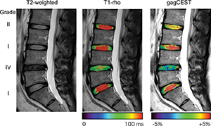
Low back pain (LBP) is a leading cause of disability worldwide.1 The most common cause is mechanical, and although most individuals recover in a short amount of time with minimal intervention, recurrences are common, and some will even go on to experience “chronic” pain, or pain lasting longer than 3 months. It is often difficult to isolate an anatomic correlate of LBP because a number of structures in the lumbar spine, including, but not limited to, vertebrae, discs, facet joints, muscles, and ligaments, may serve as active pain generators.2-4 However, it is important to note that such a determination is typically not necessary, as the primary treatment of uncomplicated LBP is usually conservative and aimed at symptom alleviation.
Routine imaging in LBP is not recommended, given that modalities often have false-positive and false-negative results. Imaging in acute LBP has not been shown to yield new, significant findings or alter outcomes5-6 and its role in chronic LBP is even more dubious. Previous studies have shown that a large proportion of individuals without LBP have disc herniations.7-9 In addition, a study by Savage et al10 revealed that nearly a third of asymptomatic patients had abnormal lumbar spine findings characterized by disc degeneration, disc bulging, facet hypertrophy, or nerve root compression, and that less than half of patients with back pain had an identifiable abnormality. The American College of Physicians currently recommends imaging for severe, progressive neurologic deficits, or in the setting of certain “red flags,” such as new urinary retention or overflow incontinence, a history of cancer, a recent invasive spinal procedure, and significant trauma, as well as in those who fail conservative treatment.

