Warning: Declaration of My_Walker::start_el(&$output, $item, $depth, $args) should be compatible with Walker_Nav_Menu::start_el(&$output, $data_object, $depth = 0, $args = NULL, $current_object_id = 0) in /home2/ajnrblog/public_html/ajnrdigest/wp-content/themes/ajnr/functions.php on line 258
Mild Traumatic Brain Injury - AJNR News Digest
March 2014
Introduction

Mild Traumatic Brain Injury

Saeed Fakhran

Saeed Fakhran

The imaging of mild traumatic brain injury (MTBI), commonly referred to as concussion, is a maturing and expanding field, with novel imaging methods being used for both clinical evaluation of patients as well as in research. While initial concussion imaging was focused on excluding more serious pathology (eg, hemorrhage) in these patients, the field has long since moved beyond this rather simple start. Subsequent work has focused on evaluation of abnormalities on DTI, which serve as markers of microstructural changes implicated in diffuse axonal injury. This evaluation has focused predominantly on detecting these DTI white matter changes in patients with MTBI compared with controls, with multiple studies demonstrating widespread abnormalities.

The challenge, thus far, has lain in making sense of these abnormalities and attempting to correlate them with either meaningful patient deficits or patient outcome. Traditionally, clinicians and radiologists have viewed concussion through a holistic lens, and thus have attempted to correlate—with largely frustrating results—patient outcome with cognitive metrics such as the Mini-Mental State Examination (MMSE).

At the University of Pittsburgh we are advancing a more nuanced approach to MTBI. Setting aside the traditional holistic approach to concussion, we instead attempt to classify concussion patients based on one of six dominant symptom clusters—anxiety, cervicalgia, sleep-wake disturbance, vestibulopathy, migraines, and ocular dysfunction—and subsequently attempt to correlate abnormalities in these subgroups
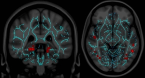
not with cognitive metrics but, instead, with patient-reported symptoms. Preliminary results, such as the association of parahippocampal abnormalities with sleep-wake disturbances, have shown an exciting and revealing structure-function relationship that has not been reported previously.

In the current issue of AJNR News Digest, we highlight the work performed in MTBI research that has furthered our understanding of the imaging appearance of white matter injury patterns underlying MTBI.

One article, by Mukherjee et al, compares the value of DTI imaging to conventional T2*-weighted gradient-echo images for detecting diffuse axonal injury in patients with MTBI that correlates with symptoms. This article nicely highlights the important role DTI plays in the evaluation of these patients, and the significance of abnormalities that are detected solely by this method.

The next two articles, by Chu et al and Wada et al, evaluated whole-brain DTI imaging in patients with MTBI using a voxel-based analysis. They described multiple areas of DTI abnormalities in areas of normal appearing white matter (by conventional MR). Furthermore, they correlated the overall degree of abnormality with the severity of postconcussive symptoms, and cognitive markers such as the MMSE.

The final series of articles, explores alternate imaging strategies in the evaluation of patients with MTBI. Different authors focus on exploring the roles of MR spectroscopy, diffusion kurtosis imaging, and brain iron quantification in the pathophysiology of MTBI.


Image modified from: Fakhran S, Yaeger K, Alhilali L. Symptomatic White Matter Changes in Mild Traumatic Brain Injury Resemble Pathologic Features of Early Alzheimer Dementia. Radiology 2013;269:249–57. doi: 10.1148/radiol.13122343