Warning: Declaration of My_Walker::start_el(&$output, $item, $depth, $args) should be compatible with Walker_Nav_Menu::start_el(&$output, $data_object, $depth = 0, $args = NULL, $current_object_id = 0) in /home2/ajnrblog/public_html/ajnrdigest/wp-content/themes/ajnr/functions.php on line 258
PET-CT Imaging in Head and Neck Cancer - AJNR News Digest
February 2015
Introduction

PET-CT Imaging in Head and Neck Cancer

Saeed Fakhran

Saeed Fakhran

Carcinoma of the squamous lining of the mucosal surfaces of the head and neck, collectively known as head and neck cancer, accounts for over 50,000 cases per year and represents approximately 3% of all cancers in the United States. Traditionally, head and neck cancer was thought to be a disease of older people (males greater than females) with a history of alcohol and tobacco abuse. With recent increasing awareness of the risks of oral HPV infection, there is growing concern and realization that head and neck cancer is no longer a disease of the elderly.

Since its widespread introduction in 2001, the hybrid technique of PET/CT imaging has gained an ever increasing foothold in oncologic imaging. It is now widely used in the initial diagnosis, staging, assessment of therapy response, and long-term surveillance of a wide array of tumors throughout the body. Predictably, PET/CT has also gained traction as the dominant modality used to diagnose, treat, and monitor therapy response in head and neck squamous cell carcinoma. It goes without saying that a basic familiarity with PET imaging, artifacts, and expected normal findings within both the neck and the rest of the body is required to provide a clinically meaningful interpretation.

There still exists significant between-institution variability in both the protocols of PET/CT studies (with or without intravenous contrast, with low-dose CT technique or a diagnostic quality CT) and the frequency of surveillance of PET/CT studies in the management of head and neck cancer. As of now, no evidence-based recommendations in these areas exist.
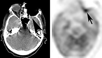
In this issue of AJNR News Digest, we focus on the work of authors who shed light on the optimal methods for use of PET/CT in head and neck cancer. An excellent review article by Srinivasan et al discusses the various uses of PET/CT in imaging of head and neck cancer, and highlight novel uses of biologic imaging (including PET/CT) in the current and future imaging of head and neck cancer.

Hughes et al discuss optimization of surveillance PET/CT in treated head and neck squamous cell carcinoma. They found that while an initial negative PET/CT has a high negative predictive value, 2 consecutive negative PET/CT studies in a 6-month period is required to halt further imaging surveillance. Appropriate use of imaging in cancer surveillance is an area of increasing importance, in light of the current focus on streamlining imaging costs and economizing the use of health care dollars.

The next article, by Hoang et al, discusses the use of intratreatment change in standard uptake values (SUV)  to determine early treatment response (or lack thereof) in head and neck squamous cell carcinoma, given baseline variability of SUV on FDG-PET scans. They found that treatment related SUV changes exceeded variability of baseline SUV values in nodal disease and can be used to assess treatment response.

Despite the metabolic information that PET/CT adds to oncologic imaging, it is not superior to conventional anatomic imaging in all situations in which there is a question of tumor recurrence. The final article, by Alhilali et al, discusses the relative failure of SUV values on PET/CT to help distinguish osteoradionecrosis from recurrent head and neck carcinoma, compared with conventional CT imaging findings.


Image modified from: Srinivasan A, Mohan S, Mukherji SK. Biologic imaging of head and neck cancer: the present and the future.