Benjamin M. Ellingson
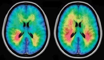
Investigators from our team recently discovered that patients with glioma having mutation of the isocitrate dehydrogenase-1 (IDH1) gene tended to have larger, nonenhancing tumors localized in the frontal lobe (Lai et al, J Clin Oncol, 2011; 29:4482-90). Using an approach similar to lesion-symptom mapping, we developed a technique where MR images of the brains of patients with glioma were registered to stereotactic space, their tumors contoured, and the frequency of tumor occurrence spatially mapped and statistically compared between IDH1 mutant and wild type tumors (Analysis of Differential Involvement, ADIFFI maps). Results clearly suggested these tumors likely arise from a very specific region in the frontal lobe. Based on this interesting observation we decided to examine whether tumors with O(6)-methylguanine-DNA methyltransferase (MGMT) promoter methylation tended to occur more frequently in a specific location in the brain (Ellingson et al, Neuroimage, 2012; 59:908-16). Interestingly, we noticed that most tumors tended to be contiguous with the subventricular zone, known to harbor adult stem cells, and MGMT promoter methylated tumors tended to be lateralized to the left temporal lobe. Additionally, we noticed that if patients had tumors near this specific region in the left temporal lobe they were more likely to live longer, independent of their methylation status. At this time the University of California Cancer Research Coordinating Committee (UC CRCC) decided to help us build the first set of Probabilistic Radiographic Atlases for various tumor genotypes and phenotypes, comparing tumor volumes and
