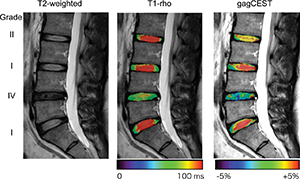
Degeneration of intervertebral discs (IVDs) is one of the leading causes of low back pain.1 Surgical treatment has been performed in severe IVD degeneration; however, early-stage IVD degeneration could be treated with regenerative medicine therapy.2 Therefore, noninvasive and quantitative imaging methods to detect and monitor changes of IVD degeneration are desirable. Proteoglycans (PGs) and glycosaminoglycans (GAGs) are the platform of the cartilage matrix, and they play crucial roles in the function of IVDs. So far, the Pfirrmann grade is widely used to qualitatively asses IVD degeneration on T2-weighted images. However, T2-weighted imaging cannot detect the loss of PGs or GAGs. It has been reported that the loss of PGs can be detected by T1-ρ measurements.
However, the clinical applicability of T1-ρ imaging is limited by the long scan time and the high specific absorption rate required by multiple and long spin-lock pulses.
Chemical exchange saturation transfer (CEST) imaging has drawn considerable attention in the field of molecular imaging as a novel contrast mechanism in MR imaging.3


