Erin Simon Schwartz
While diffusion imaging of the brain parenchyma has been in widespread clinical use for nearly 20 years, use of diffusion tensor imaging of the spinal cord has been less enthusiastically embraced by the neuroradiology community. However, this technique can add valuable quantitative information to more standard imaging sequences in a variety of conditions affecting the spinal cord, particularly the cervical spinal cord, including demyelinating diseases, inflammatory myelopathies, spondylotic myelopathies, and scoliosis.
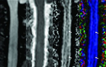
In the first paper featured in this AJNR Digest, Hesseltine, Law, and colleagues utilized DTI in a series of patients with relapsing-remitting multiple sclerosis, to assess regions of the cervical spinal cord that appeared unaffected on conventional imaging sequences. Numerous DTI metrics in their subjects were significantly different from controls, with high sensitivity and specificity, suggesting that DTI could be used to detect otherwise occult disease and provide a quantitative measure of both disease progression and response to treatment.
In the developing spine, DTI has been shown to be useful in the assessment of adolescent idiopathic scoliosis. In the cohort studied by Kong, Chu, Wang, and colleagues, changes in white matter integrity were present in the upper and mid portions of the cervical spine, as well as the medulla oblongata. They hypothesized that significant changes in fractional anisotropy (FA) and mean diffusivity (MD), and an increased incidence of cerebellar tonsillar ectopia, reflect neurophysiologic changes related to subclinical spinal cord tethering from asynchronous osseous and neural growth in this patient population. DTI could well serve as a quantitative method for pre- and posttreatment evaluation, and even for determining treatment options themselves.
