Warning: Declaration of My_Walker::start_el(&$output, $item, $depth, $args) should be compatible with Walker_Nav_Menu::start_el(&$output, $data_object, $depth = 0, $args = NULL, $current_object_id = 0) in /home2/ajnrblog/public_html/ajnrdigest/wp-content/themes/ajnr/functions.php on line 258
Susceptibility-Diffusion Mismatch Predicts Thrombolytic Outcomes: A Retrospective Cohort Study - AJNR News Digest
May 2015
Brain

Susceptibility-Diffusion Mismatch Predicts Thrombolytic Outcomes: A Retrospective Cohort Study

Zhicai Chen

Zhicai Chen

JianZhong Sun

JianZhong Sun

Intravenous thrombolysis with recombinant tissue plasminogen activator is a proved treatment for acute ischemic stroke within 4.5 hours of symptom onset. The foundation of intravenous thrombolysis for ischemic stroke involves reperfusion of the ischemic penumbra and salvage of threatened but potentially viable brain tissue. Work on imaging of the penumbra and clinical selection using penumbral evaluation have been one of the major focuses of our group. Despite the promise offered by PWI and DWI MR studies to advance patient selection for reperfusion therapies beyond noncontrast CT, sensitivity and specificity were still low. There is no consensus about the optimal perfusion parameter to accurately define ischemic tissue, and it is uncertain whether perfusion lesion volumes overestimate the penumbra tissue. So, there remains a thirst to develop new techniques that may potentially represent the penumbra based on the cerebral pathophysiology of an individual patient.

Recently, the use of blood oxygen level-dependent (BOLD) imaging as an alternative to DWI-PWI mismatch has stirred a lot of interest. It is sensitive to an increased concentration of deoxyhemoglobin (DHb) and may therefore be an indirect marker of oxygen metabolism. There are two approaches to
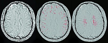
detect an increased oxygen extraction fraction (OEF) using BOLD: 1) assessment of tissue and 2) assessment of draining veins containing an increased fraction of DHb. SWI was used in clinical practice to exclude hemorrhage before intravenous thrombolysis, and it allows clear visualization of cerebral veins. That’s why we chose this topic: we believed SWI venography may provide the oxygen metabolic information about ischemic brain tissue by noninvasive estimation of blood oxygen level.

In our research we found that patients with prominent asymmetry of cerebral veins on SWI were more likely to have severe arterial occlusion and hypoperfusion, and patients with prominent asymmetry of cerebral veins on SWI and a relatively small diffusion-weighted imaging lesion were more likely to benefit from thrombolytic therapy. As a result of this study, we have implemented this concept for patient selection in intravenous thrombolysis beyond 4.5 hours.

Our group is going to continue the research in cerebral veins on SWI. We are testing the application of cerebral veins in different locations, trying to find some key veins in acute patients with ischemic stroke. We are also trying to provide a simple and accurate assessment measure for clinical physicians.


Read this article at AJNR.org …