 Winnie Chu |
 Defend Wang |
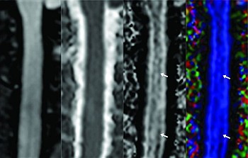
This topic was chosen because our group has a long history of research interest in the etiopathogenesis of adolescent idiopathic scoliosis (AIS). In our previous work, which evaluated the morphologic aspect of the spinal cord in AIS using MRI, we found that patients with AIS had increased total vertebral column length, predominantly affecting the thoracic segment, without corresponding lengthening of the spinal cord; hence, there is a reduced cord-vertebral length ratio in AIS, associated with distortion of cross-sectional shape of the cord at the level of scoliotic apex and resulting low-lying cerebellar tonsils. The above observations support the hypothesis of asynchronous or uncoupled neuro-osseous growth, first proposed by Roth and rediscovered by Porter. It is hypothesized that when spurts of elongation of the spine are too rapid for the slower growth rate of the spinal cord and nerve roots, an abnormal rotator anatomy is observed at the apex in scoliosis. As a result, lordosis and adaptive scoliotic curvature develop in the otherwise normally growing spine. According to the above theory, the spinal cord might fail to stretch in response to vertebral growth, and there will be damage to the white matter tract at the molecular level. Findings of our current study provide further support to the above theory. By using advanced diffusion tensor imaging, we have demonstrated significantly decreased fractional anisotropy (FA) values and increased mean diffusivity (MD) values at the medulla oblongata and upper cervical segment (C1–C5) of the spinal cord in patients with AIS when compared with healthy controls.